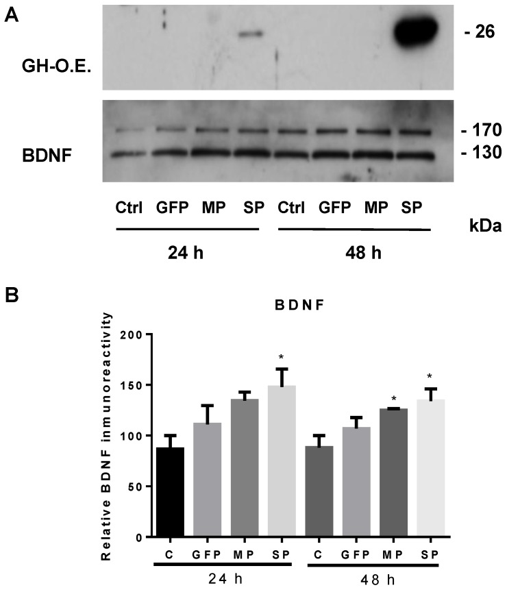
Figure 2.
Effects of growth hormone (GH) over-expression on brain-derived neurotrophic factor (BDNF) secretion in neuroretina-derived quail cell line (QNR/D) cell cultures. Culture media (20 µL) were analyzed at 24 and 48 h post-transfection by Western blot (reducing conditions). (A) Representative luminograms of GH (top) and BDNF (bottom) in the culture media; (B) relative changes in BDNF immunoreactivity were determined by densitometry (n = 3). Control (C; not transfected) and green fluorescent protein (GFP; pCAG plasmid by Addgene) overexpression groups were used as controls. MP (mature peptide) is a plasmid construction expressing a non-secreted GH and the SP (signal peptide) construction that produces a secreted GH. Transfections were performed as reported in [71]. Asterisks show significant differences (p < 0.05) in comparison to the control (C), as determined by one-way ANOVA and Tukey post hoc test.

