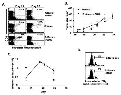
Figure 2.
Anti-CD40 treatment accelerates the deletion of tumor-specific T cells. Mice were challenged with 1 × 10⁵ B16ova cells intradermally. Seven days later, when the tumor was palpable, mice were injected with 200 μg of either anti-CD40 or control antibody i.p. (A) At the times indicated, the tumor was removed, stained, and analyzed as described in the legend of Fig. 1. (B and C) Tumor-bearing mice were injected with 200 μg of anti-CD40 i.p. on day 7 after initial tumor challenge and every 7 days thereafter (i.e., days 14 and 21). The data were analyzed as described in the legend of Fig. 1C. (D) Fifteen days after initial tumor challenge and 8 days after anti-CD40 treatment, cells from the tumors of control and anti-CD40-treated mice were stained as described in Materials and Methods for intracellular IFNγ. The data shown (solid histograms) have been gated on all class II−, CD8+, tetramer+ events. The background (open histogram) is from gating on all non-tetramer-staining CD8+ T cells in the tumor. These results are representative of three separate experiments.

