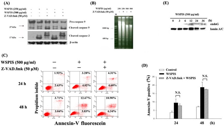
Figure 5.
Caspase-independent apoptosis in WSPIS-treated THP-1 cells. (A–D) Cells were pretreated with or without Z-VAD.fmk (50 μM) for 1 h followed by WSPIS treatment for 24 and 48 h. (A) Cleaved products of caspase-3 and caspase-9 (17 and 37 kDa, respectively) were measured by Western blotting. (B) Oligonucleosomal DNA fragmentation was determined by agarose gel electrophoresis. M is the DNA markers. (C) Apoptotic cells (annexin-V-positive) were analyzed using flow cytometry after annexin-V/propidium iodide staining. (D) Apoptotic cells are expressed as mean ± SD of three independent experiments. N.S. = Not significant. (E) Nuclear translocation of endoG. Cells were treated with 500 μg/mL WSPIS for the indicated time periods. The levels of nuclear endoG were determined by Western blotting. Results are representative of three independent experiments.

