Figure 7.
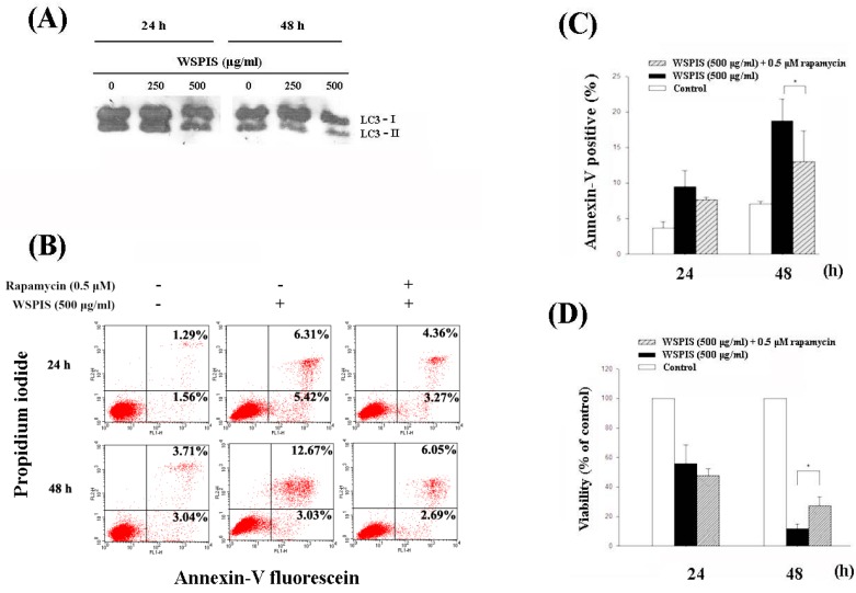
WSPIS inhibited autophagy. (A) Cells were treated with various concentrations of WSPIS (250 and 500 μg/mL) for 24 h and 48 h. The protein levels of LC3-I and LC3-II were measured by Western blotting. (B) Autophagy induction by rapamycin partially rescued WSPIS-induced apoptosis. THP-1 cells were pretreated with rapamycin (0.5 μM) for 1 h, followed by 500 µg/mL WSPIS treatment for 24 and 48 h. Apoptotic cells were analyzed by using flow cytometry after annexin-V/propidium iodide staining. (C,D) Rapamycin partially rescued WSPIS-induced apoptosis and cell death after 48 h treatment. Apoptosis and cell viability are expressed as mean ± SD of three independent experiments. Cell viability was determined by trypan blue staining. * denotes the data that are significantly different between WSPIS-only and WSPIS-combined rapamycin groups at p < 0.05.

