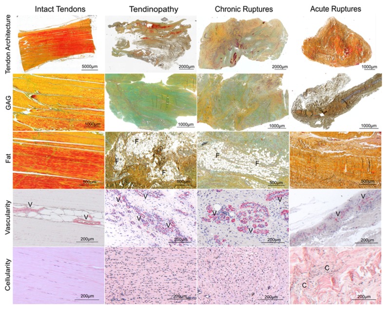
Figure 1.
Exemplary histologies of intact tendons, tendinopathic tendons, chronic ruptures and acute ruptures using Movat Pentachrome (MP) staining (tendon architecture, glycosaminoglycan (GAG) and fat tissue (lipid vacuoles)), α smooth muscle actin (αSMA) staining (vascularity) and Hematoxylin Eosin (HE) staining (cellularity). F: fat tissue, V: vessels, C: cell clusters. The Movat Pentachrome staining used stains collagen in yellow to brown, mature collagen in red [31], GAG ground substance in turquois, cell nuclei in blue to black and cytoplasm stains reddish.

