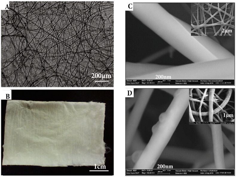
Figure 8.
(A) Macroscopic image of chitosan fibre and (B) fibrous mat; (C) Morphology of fibre evaluated by SEM and atomic force microscopy of 0.1% genipin crosslinked and 1% HA loaded; (D) 7% chitosan fibres, typical morphology seen inset images [143]; (Adapted with permission of publisher).

