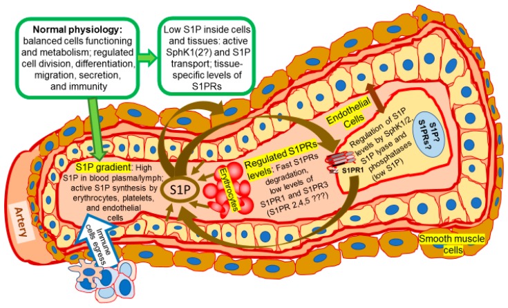
Figure 1.
Normal physiology: the place of sphingolipid signaling pathway. A transverse cut through the artery shows an inside space of the blood vessel filled with blood plasma and blood cells (only erythrocytes are shown), as well as a layer of endothelial cells surrounded by smooth muscle cells. Endothelial cell layer and enlarged single endothelial cell are indicated by a double-ended arrow. Coordinated sphingolipid signaling creates a favorable environment for normal physiological functioning. The activity and expression levels of sphingosine kinases (SphK), sphingosine-1-phosphate (S1P) and its receptors (S1PRs) are maintained at normally low tissue specific levels required for healthy metabolism. Alternatively, high concentration of S1P in blood plasma (and lymph) is supported by release of S1P from endothelial cells, erythrocytes, and platelets. Binding of S1P to S1PRs stimulates activation of an appropriate signaling in cytoplasm and gene activation in nucleus that are followed by quick S1PRs internalization, degradation, and re-cycling. Activation of endogenous SphK by hormones, cytokines, and growth factors results in reduction of sphingosine cell content and production of S1P. S1P/S1PRs axis activates further downstream signaling targets and controls variety of physiological processes including lymphocyte egress from lymphoid organs [5,8,9,14,17,21,62,63,64,65,66,67,68]. Arrows indicate the movement of S1P and direction of its effects (such as activation of S1PR by S1P as ligand, release of S1P by endothelial or cancer cells, etc.). The arrow directed from S1P to the endothelial cell indicates that S1P also activates endothelial cell signaling via S1PRs. Question marks indicate the gaps in our knowledge of SphK/S1P/S1PRs axis.

