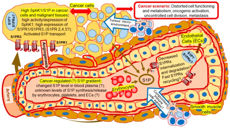
Figure 2.
Cancer scenario: Transformation of SphK/S1P/S1PRs signaling. Cancer cells are shown outside of the artery wall. A transverse cut through the artery shows an inside space of the blood vessel filled with blood plasma and blood cells (only erythrocytes are shown), as well as a layer of endothelial cells surrounded by smooth muscle cells. Endothelial cell layer and enlarged single endothelial cell are indicated by a double-ended arrow. Levels of SphK1 expression and activation are high in cancer cells. The regulation of plasma S1P levels and S1P release by blood cells/endothelial cells remain to be confirmed in cancer patients. S1P levels/S1P gradient is hypothetically changed in blood plasma of cancer patients comparing to healthy controls. It is not clear how changed S1P plasma level would influence the level of S1PR expression and signaling. Overactivation and delayed degradation of S1PRs in cancer cells results in reinforced signaling of this pathway and cancer-related pathological consequences including cancer-directed angiogenesis [21,22,23,70,71,72,73,74,75], formation of tumor microenvironment [32,76,77,78], transformed metabolism [68,79], and cancer progression [5,6,11,67,80,81,82,83]. Arrows indicate the movement of S1P and direction of its effects. The arrow directed from S1P to the endothelial cell indicates that S1P also activates endothelial cell signaling via S1PRs. Question marks indicate the gaps in our knowledge of SphK/S1P/S1PRs axis.

