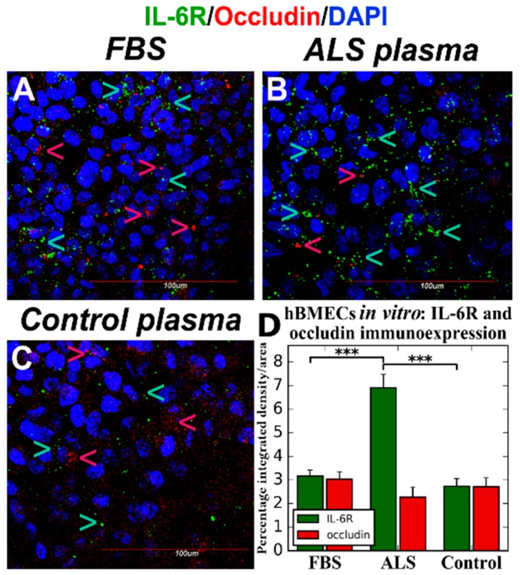
Figure 1.
Confocal fluorescent images of hBMECs immunostained for IL-6R and occludin in vitro. Double immunostaining for IL-6R and occludin was performed on fixed hBMECs after 5 DIVexposure to FBS, plasma from ALS or control patient. The cells containing ALS plasma in media demonstrated significantly increased IL-6R (green, arrow) and reduced occludin (red, arrow) immunoexpressions (B,D). There were no differences in IL-6R or occludin immunoexpression between culture cells after exposure to FBS (A) or plasma from control subject (C). DAPI (blue) was used for nuclei staining. Data are presented as means ± S.E.M. Scale bar in (A–C) is 100 µm. *** p < 0.001.

