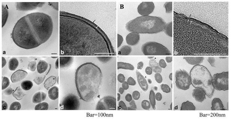
Figure 1.
Internal morphology of S. aureus (A) and E. coli (B) observed via TEM (a,b) untreated bacteria, (c,d) bacteria treated with Ag+ (0.2 ppm) during 2 h. Black and white arrows indicate peptidoglycan and cytoplasmic membrane, respectively (A) and outer membrane, peptidoglycan and cytoplasmic membrane (B). Arrowhead indicate separation of the cell membrane from the cell wall. Reprinted from [40] with American Society for Microbiology Publishing Group permission.

