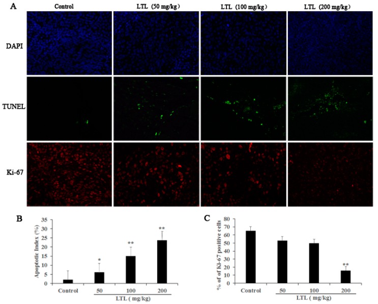
Figure 4.
The effect of LTL on tumor cell apoptosis and proliferation in vivo. Paraffin sections of tumor tissue were tested by TUNEL (terminal deoxynucleotidyl transferase dUTP nick end labeling) and Ki-67 staining analysis. (A) TUNEL-positive cells (green) and Ki-67-positive cells (red) were observed under a fluorescence microscope (×400). Nuclei were counter-stained with DAPI (blue); (B) The apoptotic index was calculated as the number of TUNEL-positive cells for each group; (C) Quantification of Ki-67-positive cells is represented as the ratio of Ki-67-positive cells to the total number of cells for each group. The results are expressed as means ± SD of three independent experiments. * p < 0.05, ** p < 0.01, compared with the controls.

