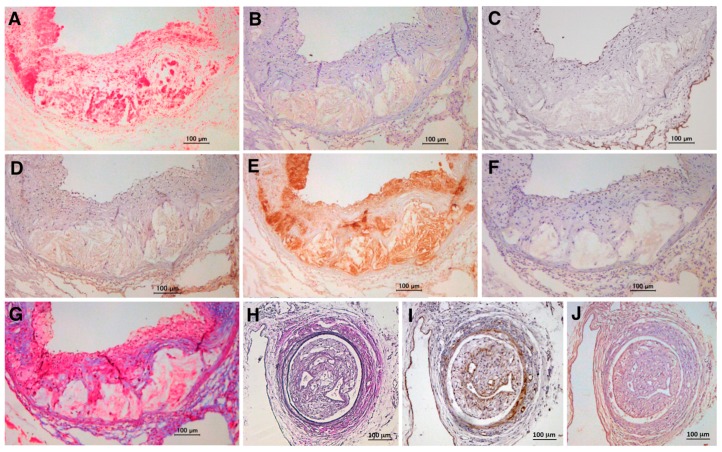
Figure 4.
Expression of TSG-6 in atherosclerotic and restenotic lesions in ApoE-deficient mice. TSG-6 is expressed at high levels in aortic atherosclerotic lesions (A–G) and femoral artery restenotic lesions after wire injury (H–J) in ApoE-deficient mice fed a high-cholesterol diet. Tissues were immunostained with Oil Red O (Wako Pure Chemical Industries, Osaka, Japan; (A)); anti-TSG-6 antibody (Bioworld Technology, St. Louis Park, MN, USA; (B,J)); anti-podocalyxin antibody (Life Technologies, Carlsbad, CA, USA; (C)); anti-pentraxin-3 antibody (Bioss Antibodies, Woburn, MA, USA; (D)); anti-MOMA-2 antibody (Millipore, Billerica, MA, USA; (E)); anti-α-SMA antibody (Sigma, St. Louis, MO, USA; (F,I)); Masson’s Trichrome (Muto Pure Chemicals, Tokyo, Japan; (G)); and Elastica-Van Gieson (Muto Pure Chemicals; (H)). Hematoxylin was used for nuclear staining. Bar = 100 μm. (C) Podocalyxin is used as an EC marker. (G) Masson’s Trichrome is used to stain collagen fibers in blue. Panels (A–G) show unpublished data from our previous experiments [10], and panels (H–J) show unpublished data from our new experiments.

