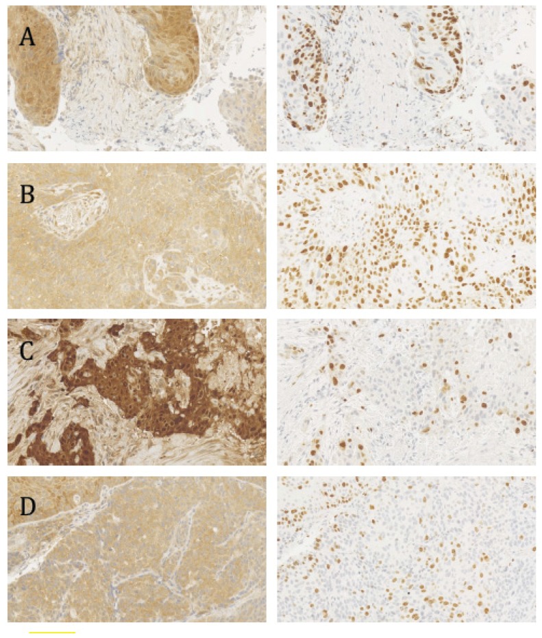
Figure 1.
p16INK4a and Ki-67 staining (left and right panels, respectively) in consecutive sections of human papillomavirus (HPV)-positive and HPV-negative tumor biopsies. (A) HPV-positive, diffuse nuclear p16INK4a staining; (B) HPV-positive, diffuse cytoplasmatic p16INK4a staining; (C) HPV-negative, diffuse nuclear p16INK4a staining. (D) HPV-negative, diffuse cytoplasmatic p16INK4a staining. The proliferation index (Ki-67 staining) is significantly higher in HPV-positive tumors. (A–D) were obtained with 20× magnification.

