Figure 2.
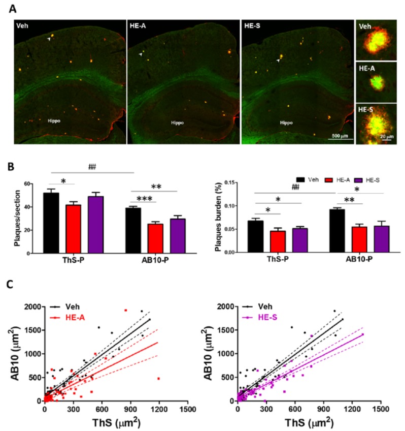
HE-A and HE-S reduce amyloid plaque burden and size in APP/PS1 mice. APP/PS1 transgenic mice orally administered with vehicle (Veh) and HE-A or HE-S for 30 days (n = 8 for each group), and then amyloid plaques were stained by thioflavin S (ThS) and immuno-stained with AB10 antibody. (A) The representative fluorescent images of ThS (green) and AB10 (red) in the indicated area were shown. Hippo, hippocampus. Scale bar: 500 μm. The typical plaques (arrow) are magnified and is shown at the right side. Sale bar: 20 μm; (B) The number and burden of ThS- stained plaque (ThS-P) and AB10-stained plaque (AB10-P) in cerebral hemisphere were calculated by image analysis software. Plaque burden is displayed as a percentage of the area occupied by ThS or AB-10 stained signal in the full area of interest. The results are the mean ± standard error of mean (S.E.M). Significant differences between Veh group and the other groups are indicated by *, p < 0.05; **, p < 0.01; ***, p < 0.001. Significant differences between 30 d-ThS group and the other groups are indicated by ##, p < 0.01; (C) Scatter plots of ThS- and AB10-stained area of each single plaque in the brain slice. (Solid lines: linear regression lines; dashed lines: 95% confidence intervals).

