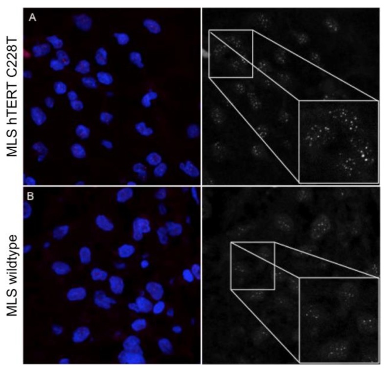
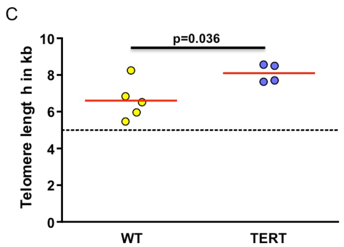
Figure 2.
Representative Q-FISH images captured by confocal laser scanning microscopy: (A) C228T TERT promoter-mutated myxoid liposarcoma, and (B) a TERT promoter wildtype myxoid liposarcoma patient. Magnification 630×. (C) Quantification of telomere length after Q-FISH analysis (arbitrary units, a.u.). The dashed line represents 5 kb, the threshold of critical short telomeres.


