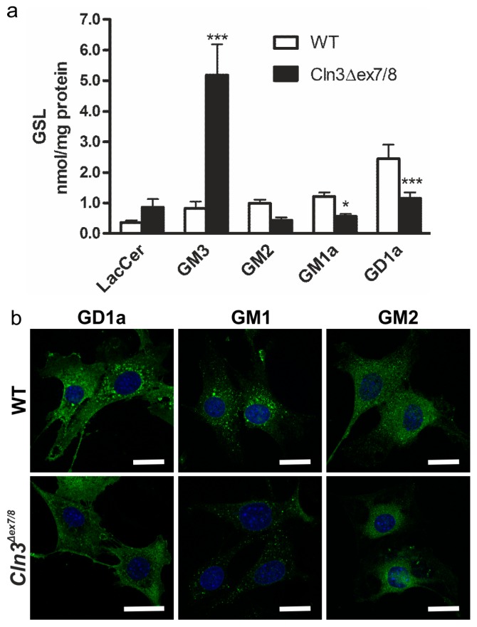
Figure 2.
Alterations in gangliosides in the cerebellar precursor cells from the Cln3Δex7/8 mouse model. (a) Quantitative HPLC analysis of glycosphingolipids in wildtype (WT) and Cln3Δex7/8 cells shows increased levels of GM3, together with a reduction in GM1a and GD1a levels. Statistical analysis with 2-way ANOVA. p values * p < 0.05; *** p < 0.001; (b) Staining of GD1a, GM1a and GM2 in WT and Cln3Δex7/8 cells. GM1a and GD1a levels are reduced in Cln3Δex7/8 cells as compared with WT cells. The cells were immunostained with specific antibodies against the respective gangliosides and detected with a secondary antibody coupled to Alexa 488. Blue staining shows the nuclei stained with DAPI. Scale bar: 20 µm.

