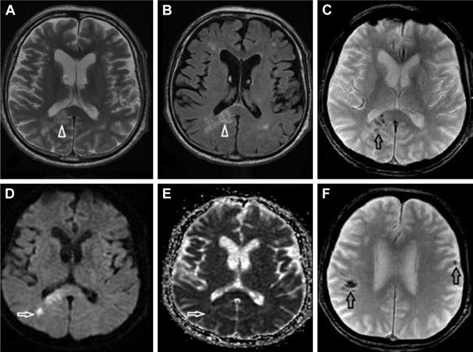
Figure 3.
SCNSL mimicking multiple ischemic lesions.
Notes: Multiple hyperintense lesions on T2-weighted images (A) and FLAIR (B) were observed (white arrowheads). The lesions showed diffusion restriction (D; white arrow) and low signal on the ADC map (E; white arrow). Nearly all lesions had signs of hemorrhage on the gradient echo sequence (C and F; black arrows).
Abbreviations: ADC, apparent diffusion coefficient; FLAIR, fluid-attenuated inversion recovery; SCNSL, secondary central nervous system lymphoma.

