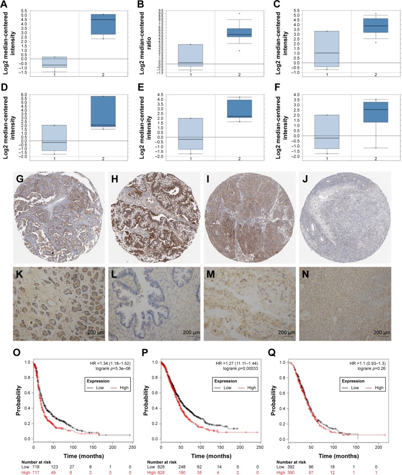
Figure 1.
Immunohistochemical analysis of SPP1 expression in epithelial ovarian cancer.
Notes: The mRNA expression of SPP1 in epithelial cancer was investigated. Data were derived from Oncomine database. (A–C) Three independent data (Adib Ovarian, Yoshihara Ovarian, and Lu Ovarian) showed that the mRNA level of SPP1 in ovarian serous adenocarcinoma was higher than in normal ovarian tissues. (D–F; data derived from Lu Ovarian) The mRNA level of SPP1 in ovarian clear cell adenocarcinoma, ovarian endometrioid adenocarcinoma, and ovarian mucinous adenocarcinoma was higher than in normal ovarian tissues. (G–I) The location and expression of SPP1 protein in ovarian cancer tissues was analyzed. The data were derived from the human protein atlas. The expression of SPP1 in ovarian serous adenocarcinoma, ovarian mucinous adenocarcinoma, and ovarian endometrioid adenocarcinoma showed moderate staining. (J) SPP1 in the follicle cells showed medium staining. SPP1 was not detected in ovarian stroma cells. (K–N) The expression pattern of SPP1 was determined in normal ovarian tissues and epithelial ovarian cancer tissue samples using IHC. Original magnification, 200×. SPP1 expression reversely correlated with survival time. Kaplan–Meier survival analysis for the relationship between survival time and SPP1 signature in ovarian cancer was performed by using the online tool (http://kmplot.com/analysis/). (O) PFS curves are plotted for ovarian cancer patients (n=1,436). (P) OS curves are plotted for ovarian cancer patients (n=1,657). (Q) PPS curves are plotted for ovarian cancer patients (n=782). p<0.05 was considered to be a statistically significant difference.
Abbreviations: HR, hazard ratio; OS, overall survival; PFS, progression-free survival; PPS, post-progression survival; SPP1, secreted phosphoprotein 1.

