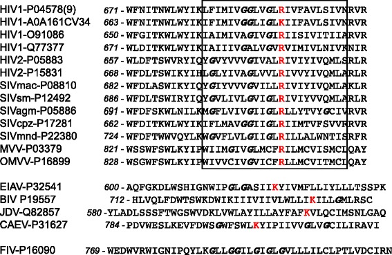
Fig. 1.

TMDs of lentiviral envelope proteins exhibit conserved charged residues. The sequence of the TMDs and surrounding region of several lentiviral envelope proteins are represented. Amino acid positions for each sequence are numbered in italic. The approximate position of the predicted TMDs is indicated. Potentially charged amino-acid residues are in red and glycine residues in italic. The Uniprot reference number of each protein is indicated
