Fig. 6.

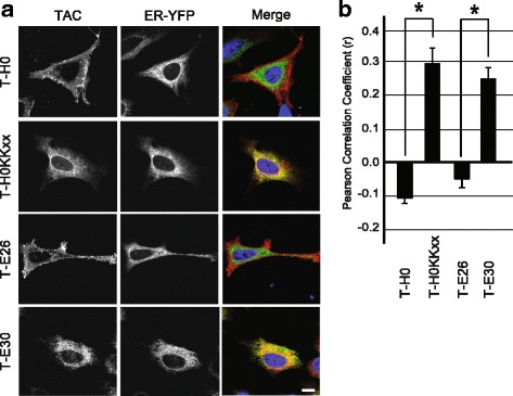
A shortened HIV-1 Env TMD acts as an ER-targeting motif. a Immunofluorescence microscopy of HeLa cells co-expressing various Tac fusion proteins (stained with an anti-Tac antibody) and a marker of the endoplasmic reticulum (ER-YFP). All pictures were taken with a confocal microscope (LSM700, Zeiss). Scale bar: 10 μm. b The colocalization of Tac proteins with the ER was quantified by measuring the Pearson’s correlation coefficient with Imaris software. T-E30 and T-H0KKxx are significantly localized in the ER (n = 4; *: Student’s t-test p < 0.01)
