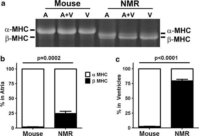
Fig. 2.

Composition of myosin heavy chain isoforms differs between mouse and naked mole-rat (NMR) in regions of the heart. a Large format SDS-PAGE electrophoresis showed α- and β-MHC in both the ventricles (V) and atria (A). Naked mole-rats had significantly more β-MHC than mice, but still expressed predominantly α-MHC in their atria. b Naked mole-rats had mostly β-MHC in their ventricles, making them more like humans (n = 11 ventricles and n = 6 atria/species)
