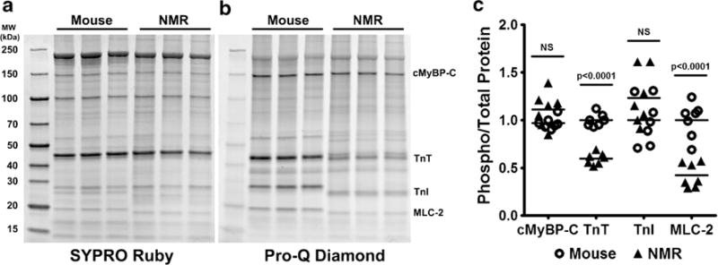
Fig. 3.

Phosphorylation of myofilament proteins is either lower or no different in naked mole-rat (NMR) ventricles when compared to those of mice. a SYPRO Ruby stain showed all myofilament fraction proteins. b Pro-Q Diamond stain showed total phosphorylation of myofilament proteins, with specific proteins highlighted: cardiac myosin binding protein C (cMyBP-C), troponin T (TnT), troponin I (TnI), and myosin light chain 2 (MLC-2). c Quantification of the highlighted proteins shows that naked mole-rats had significantly lower phosphorylation of TnT and MLC-2 than mice, but there were no species differences in cMyBP-C and TnI phosphorylation (n = 7/species)
