Fig. 2.

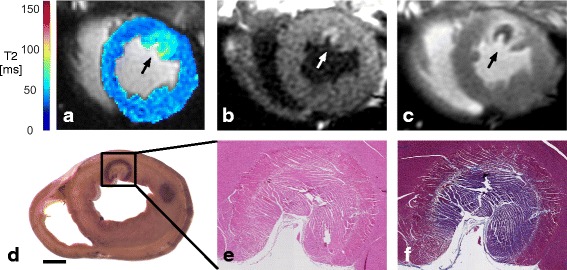
RF lesion visualization using native-contrast and contrast-enhanced CMR, gross pathology, and histopathology. a T2 map (74 min post-ablation) demonstrating T2 elevation associated with edema near the ablation site (arrow). b IR-SSFP (TI = 730 ms, 81 min post-ablation) demonstrating the hyper-enhanced lesion. c MCLE (TI = 805 ms, 106 min post-ablation, approximately 6 min post-Gd injection), demonstrating the dark region of microvascular obstruction, at the lesion centre, with bright surrounding tissue. d Gross pathology (with a second lesion slightly out of plane; scale bar = 1 cm). Magnified (e) H&E and (f) MT stained lesion tissue sections
