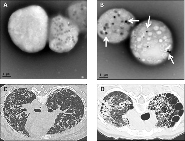
Fig 1. Electron micrograph of platelets and chest computed tomography scans in patients with Hermansky-Pudlak syndrome.
Representative whole-mount electron micrographs of platelets from a patient with Hermansky-Pudlak syndrome (A) and normal platelets (B) are shown (bar = 1 micrometer). Normal platelets contain delta granules (arrows), and platelets from patients with HPS are devoid of delta granules. Representative computed tomography scan of the chest from one patient with Hermansky-Pudlak syndrome pulmonary fibrosis showing diffuse bilateral parenchymal fibrosis with honeycombing and loss of lung volume (C) and from another patient with cystic lung destruction and upper lobe predominance of disease (D).

