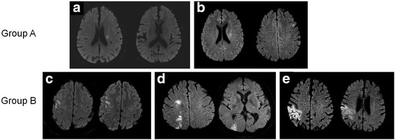
Fig. 1.

Examples of the five different diffusion-weighted imaging patterns analyzed and the classification into two groups. Lesion patterns were classified as perforating artery infarcts (a), perforating artery infarcts with additional infarcts outside the perforating artery territory (b), pial infarcts (c), border-zone infarcts (d) and territorial infarcts (e)
