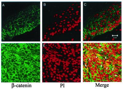
Figure 5.
Nuclear localization of β-catenin in tumors. Immunostaining and confocal image analysis shows that in WT epidermis (Upper), β-catenin is predominately localized on the plasma membranes (A), and there is no overlapping staining between β-catenin (green) and nucleus (visualized by staining with propidium iodode, red) (C). In a representative tumor sample (Lower), there is a dramatic increase of β-catenin protein in the cytoplasm (D). In addition, nuclear β-catenin can be readily detected as shown by overlapping staining (highlighted by arrows) between β-catenin (green) and nucleus (red) (F). (Scale bar = 20 μM.)

