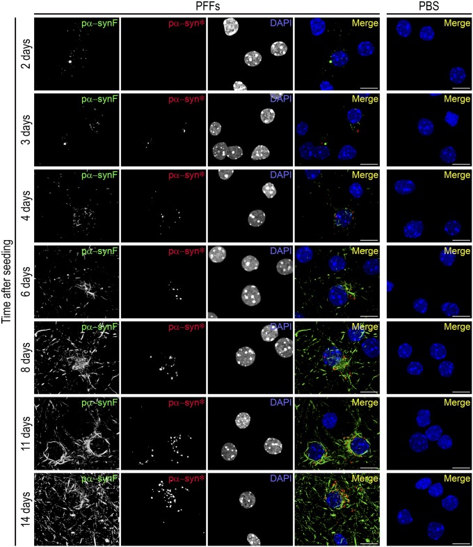
Fig. 1.
Time course of the appearance of pα-syn* in PFF-treated neurons. Primary hippocampal mouse neurons were exposed to PFFs at day in vitro (DIV)7 and were examined by immunocytochemistry (ICC) at various time points from days 2–14. Cells similarly treated with PBS alone constitute the control. Pictures show labeling with the pα-syn antibodies recognizing pα-synF or pα-syn*, respectively, and DAPI staining showing the nuclei, color-coded as green, red, and blue, respectively, in the merged image. Neurons from the PBS control were labeled similarly; the merged image is shown. No pα-syn was observed in control cells in our experimental conditions. (Scale bars, 10 µm.)

