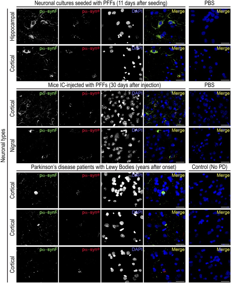
Fig. 2.
Detection of both pα-synF and pα-syn* in the brains of PFF-injected mice and PD patients. (Top) Mouse hippocampal or cortical primary neurons seeded with PFFs develop pα-synF and pα-syn* inclusions. (Middle) Mice stereotaxically injected with PFFs in the striatum develop both pα-synF and pα-syn* inclusions in the cortex and substantia nigra, with morphologies and subcellular localization identical to the cell cultures. (Bottom) Both pα-synF and pα-syn* inclusions were observed in the cortex of three LB-harboring patients with PD: case ID10-89 (Top Row); case ID12-69 (Middle Row); and case ID11-51 (Bottom Row) (Table S1). Pα-syn* was also observed in all but one of the high-LB cases and in two of the low-LB cases that we looked at. Note the pα-syn* puncta surrounding the LBs. The LBs are detected using the pα-synF–specific antibody. Labeling for pα-synF, pα-syn*, and DAPI was color-coded as green, red and blue, respectively, in the merged image. (Scale bars, 20 µm.)

