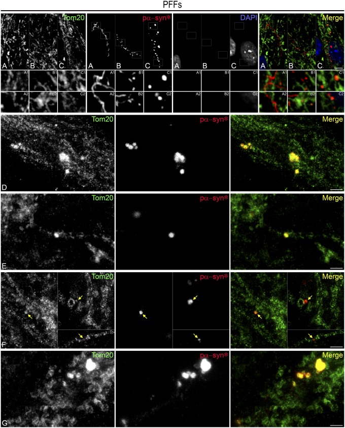
Fig. 7.
Pα-syn* localizes to mitochondria and fragmented mitochondria. (A) Immature serpentine pα-syn* is present in the vicinity of but does not colocalize with mitochondria (Insets A1 and A2). (B and C) Granular pα-syn* binds to mitochondrial tubules (Insets C1 and C2). (D–G) STED nanoscopic imaging of pα-syn* aggregates attached to mitochondrial tubules (D–F). In F, arrows point to small pα-syn* aggregates associated with mitochondrial tubules or circular structures. (G) Large pα-syn* aggregates are associated with fragmented mitochondria. Cells were labeled with Tom20 antibody (mitochondrial outer membrane) and pα-syn*, color-coded as green and red, respectively, in the merged image. [Scale bars, 5 µm in A–C; 1 µm in (D–G); Insets are a 3× magnification of the corresponding picture.]

