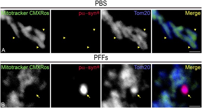
Fig. 8.
Pα-syn* induces loss of mitochondrial membrane potential. (A) Tom20 and MitoTracker CMXRos labeling in PBS-treated cells is overlapping until the ends of the tubules (arrowheads). (B) Pα-syn* labeling colocalizes with Tom20 at the end of the mitochondrial tubule, but MitoTracker CMXRos is disrupted, showing a void area (arrow). Cells were labeled with MitoTracker CMXRos, pα-syn*, and Tom20 antibody, color-coded as green, red, and blue, respectively, in the merged images. (Scale bars, 1 µm.)

