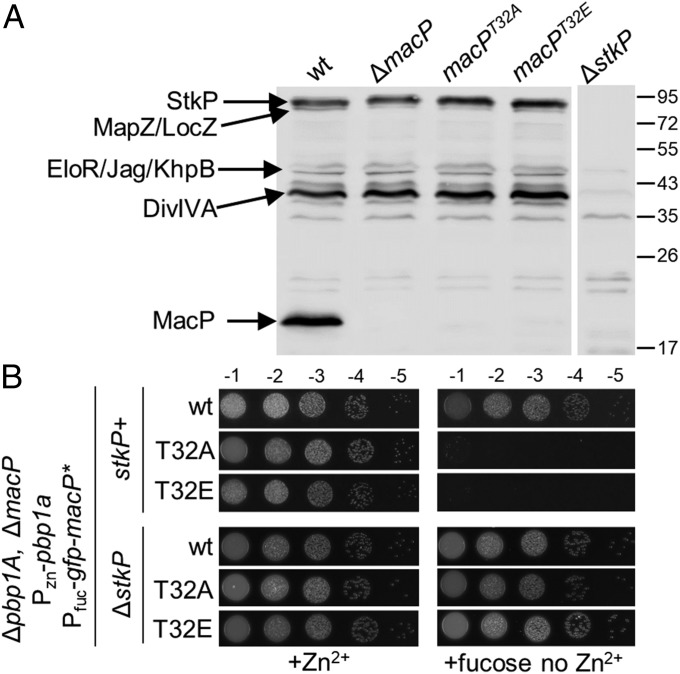
Fig. 5.
The in vivo function of MacP requires phosphorylation by StkP. (A) Antiphospho-threonine immunoblot analysis of whole-cell lysates from the indicated strains. Phosphorylated StkP and its substrates are indicated based on work from previous studies (18, 19, 29). The positions of protein markers are indicated in kiloDaltons. (B) GFP–MacP(T32A)/(T32E) do not support growth of ∆pbp1A cells depleted of MacP. The indicated strains were grown to exponential phase in the presence of 200 µM ZnCl2, normalized to an OD600 of 0.2, and 5 µL of serially dilutions were spotted onto TSAII 5%SB plates in the presence of 0.2% fucose or 200 µM ZnCl2. Plates were incubated at 37 °C in 5% CO2 and imaged. Microscopy images of lethal cell size phenotype of MacP T32A and T32E on depletion of PBP1a are shown in Fig. S5C. Growth curves of each strain are provided in Fig. S5D.

