
Figure 5. Adoptive transfer of ARE-Del−/− CD4+ T cells enhances GC responses and TBA secretion in recipient mice.
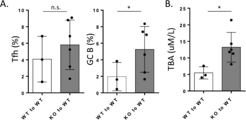
Splenic CD4+ T cells from ARE-Del−/− mice (KO) (n=6) or from control littermates (WT) (n=3) were adoptively transferred to WT mice. After 8 weeks, splenocyte populations were isolated and Tfh and GC B cells were analyzed by flow cytometry. A. Percentages of CD4+CXCR5hiPD-1hi Tfh cells and B220+CD95hiPNAhi GC B cells in spleen from KO to WT mice compared to control groups. B. Serum TBA levels were measured from KO to WT mice compared to control groups. Data represent mean ± SD. At least two independent experiments were performed. Statistical analysis was performed by the unpaired Student’s t-test. * P< 0.05, ** P< 0.01, *** P< 0.001, n.s., not significant.