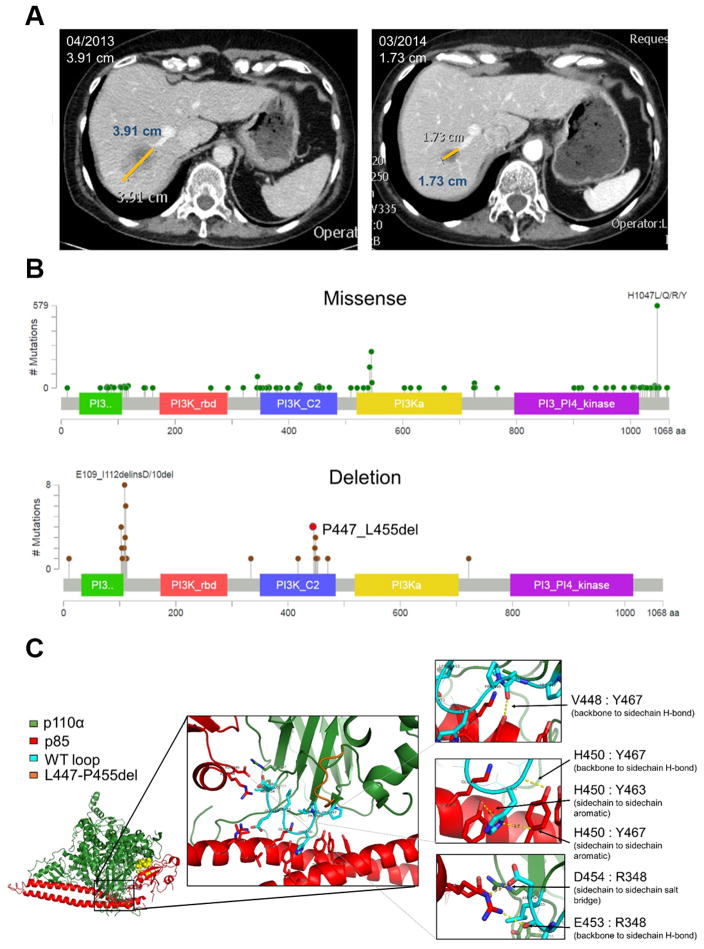
Fig. 1. PIK3CA C2 deletions occur in breast cancer and cluster predominately at p85α binding sites.
(A) Liver metastasis of patient with endocrine resistant ER+ breast cancer harboring the PIK3CAP447_L455del at baseline and 11 months after starting treatment with letrozole and alpelisib. (B) Lollipop plots of PIK3CA missense mutations (top) and deletions (bottom) from the cBioportal database (accessed 1/2017). (C) The heterodimeric structure of p110α (green) and p85α (red). Structural analysis determined the conformation of the PIK3CAWT p110α (cyan) and the interaction with p85α is altered by the deletion, PIK3CAP447-L455del (orange). Six major interaction points (arrows) are disrupted by the loss of the nine amino acids (right).

