Figure 3.

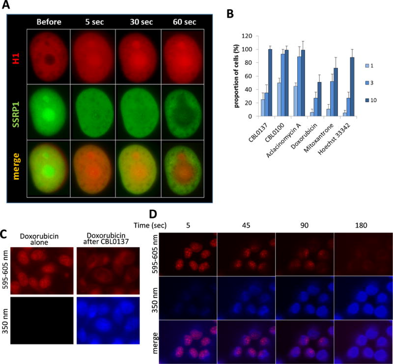
Effect of the compounds on the distribution of histone H1 in cells (A, B) and competition of curaxins and Doxorubicin for binding to DNA in cells (C and D). (A) Time-dependent effect of 3μM of CBL0137 on the distribution of mCherry tagged H1 (H1.5) and GFP-tagged SSRP1 in HT1080 cell. (B) Effect of the different doses of the compounds (in μM) on the distribution of histone H1 in HT1080 cells treated for 3 hours. Bars - proportion of cells in population with nucleoli-accumulated H1, ±SD between two experiments. (C-D) Imaging of live cells with filters corresponding to autofluorescence of the compounds: 595-605nm for Doxorubicin (red), and 350nm for curaxins (blue). HT1080 cells were incubated with 1μM of Doxorubicin for 3 hours, then 1μM of CBL0137 (C) or CBL0100 (D) was added for 10 min (C) or for the indicated times (D).
