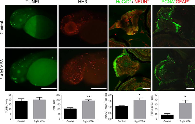
Fig. 7.

Measurement of cell death in larval neural cells by expression of TUNEL at 48 hpf, cell proliferation by HH3 staining at 48 hpf, proportion of mature newborn neurons(HuC/D+ / NEUN+ staining) at 4.5 dpf, and neural stem cell proliferation (PCNA+GFAP+ staining) at 4.5 dpf. Embryos underwent a waterborne exposure to 0.1% DMSO (control) or 5 μM VPA in 0.1% DMSO from 8hpf to 4.5 dpf. The top panel consists of representative images of each staining in controls and treatments. The bottom panel displays quantified positive cells expressions corresponding to the imagines and stains of the upper panel. n = 7-12. Values plotted are mean ± SEM. * P<0.05 and ** P<0.001 indicates significance from the vehicle control (0.1% DMSO).AC: apoptotic cells; HC: hyperplasia cells; MN: mature neuron; MAN: mitosis anaphase neuron and NSC: neural stem cells.
