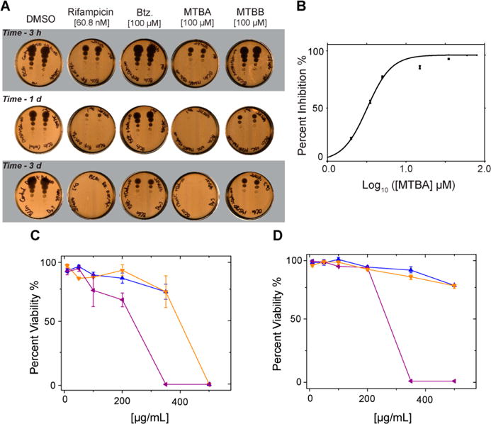
Figure 4.

Activity assay secondary screening. (A) Cell viability spot plate assay. Cells were treated with DETA-NO and either DMSO, Rifampicin, Btz, MTBA, or MTBB, for up to 6 days. Culture samples were collected at different times, diluted, plated, and incubated for 2–3 weeks prior to scanning and analysis: 3 h, 1 day, and 3 day time points are shown. (B) Inhibition of BCG cell growth under nitric oxide stress by MTBA. Colony forming unit (CFU) plate assay yields an IC50 of 8 μM. (C, D) Cytotoxic effects of MTBA (blue), MTBB (orange), and MTBC (purple) after 24 h exposure to A549 epithelial cells (C) and mouse macrophage cell line J774.16 (D).
