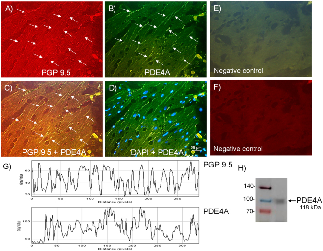
Figure 2.
Expression of PDE4A protein within nerve fibers distributed among pig bladder neck smooth muscle bundles. Double-labeling immunofluorescence assay in the pig bladder neck (A–D). Bladder neck overall innervation was visualized using the general nerve marker PGP 9.5 (red areas) (A). PDE4A immunofluorescence from pig bladder neck reveals immunopositive nerve trunks (green areas), running parallel to the smooth muscle bundles. Same fields (A,B,E and F). Immunofluorescence double-labeling for PGP 9.5 and PDE4A in the smooth muscle, demonstrate neuronal co-localization (yellow areas) (C). Cell nuclei were counterstained with DAPI (blue areas) (D). Scale bars indicate 25 µm. Negative controls showing the lack of immunoreactivity in sections incubated in the absence of the primary antibody (E and F) (n = 5). Comparison of the fluorescence of PGP 9.5 and PDE4A, using ImageJ, which shows a major co-localization between PDE4A and PGP 9.5 (G). Western blot of smooth muscle membranes from pig bladder neck incubated with PDE4A antibody showing a 118 kDa major band, which corresponded to the expected molecular weight (H) (n = 5).

