Figure 4.
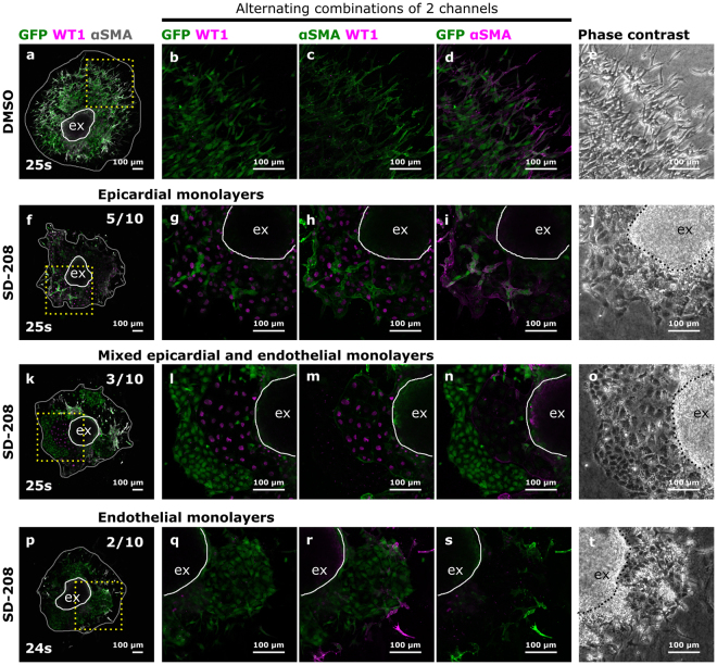
Inhibition of ALK5 leads to a relative increase in epicardial and endothelial cells. (a–t) 10 pairs of somite matched AVC explants treated with either DMSO or SD-208 in DMSO were stained for GFP, WT1 and αSMA. Boxed areas in panels (a), (f), (k) and (p) are magnified in respectively (b–d), (g–i), (l–n) and (q–s) and presented as alternating combinations of 2 channels in magenta and green. Individual confocal Z-planes are compared with the corresponding phase contrast images (e,j,o and t). The explant (ex) is delineated by a white loop. The area of cellular outgrowth in panels (a), (f), (k) and (p) is delineated by a gray loop. Upon SD-208 treatment, the observed monolayers are either epicardium only (f–j), endothelium only (p–t) or mixed epicardium and endothelium (k–o).

