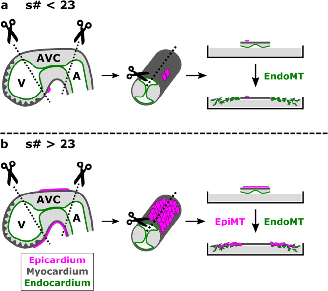
Figure 5.

Schematic representation of the AVC explant model with and without epicardial cell contamination. (a) Explants collected from embryo between 20 s–23 s contain none to nearly no epicardial cells. Only endothelium-derived mesenchymal cells will grow out of the explant. (b) From 23 s onwards, epicardial contamination increases and becomes apparent as monolayers of epicardium-derived cells capable of undergoing epicardial-to-mesenchymal transitions.
