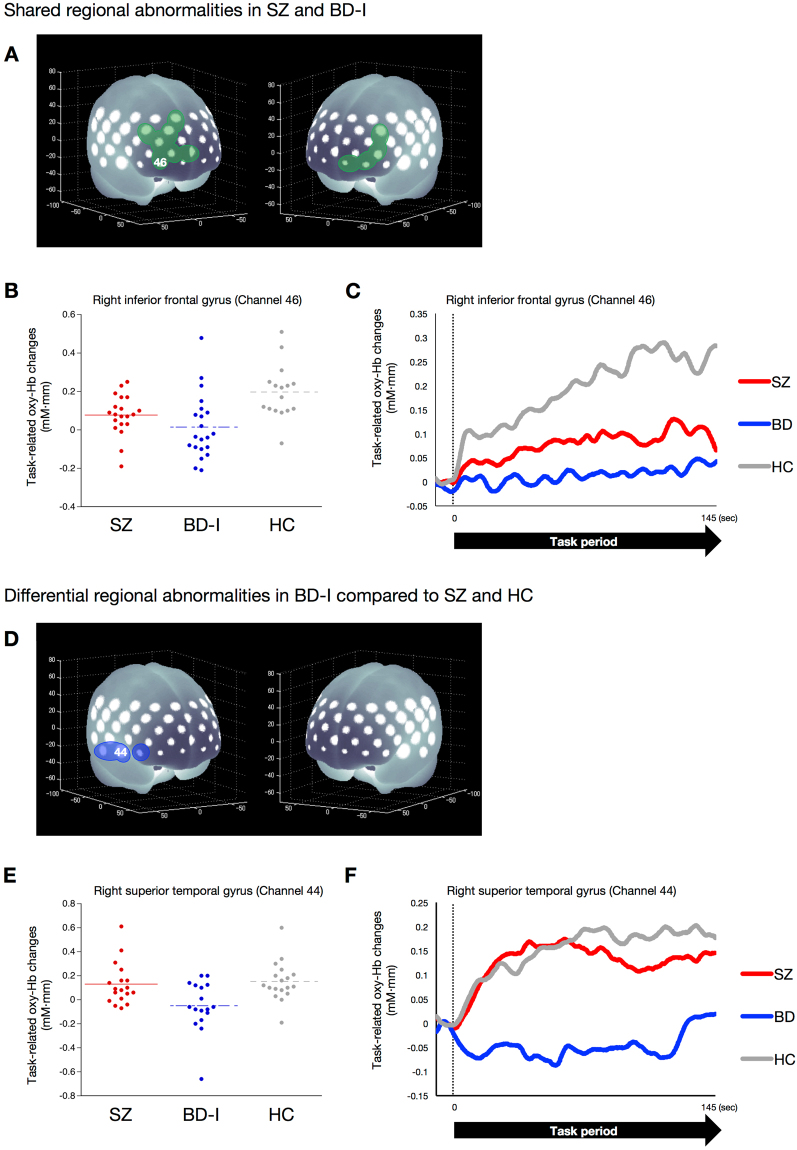
Figure 3.
Shared or differential functional abnormalities in patients with SZ or BD-I compared with HCs. (A) Brain areas in green indicate regions with reduced hemodynamic responses, compared with HCs, overlapped in both patients with SZ and BD-I: bilateral inferior frontal, middle frontal and superior frontal (including orbitofrontal region) gyri. (B) Dot plots of task-related oxy-Hb changes in the right inferior frontal gyrus (channel 46) in the SZ, BD-I and HC groups. (C) Differential time course of task-related oxy-Hb signals in the SZ, BD-I and HC groups in the right inferior frontal gyrus (channel 46). In the HC group, the time course of changes in the task-related oxy-Hb signal showed a gradual increase; the SZ and BD-I groups did not exhibit this response. (D) Brain areas in blue indicate regions where reduced hemodynamic responses, compared with SZ and HC subjects, were observed in BD-I: right inferior frontal and superior temporal gyri. (E) Dot plots of task-related oxy-Hb changes in the superior temporal gyrus (channel 44) in the SZ, BD-I and HC groups. (F) Differential time course of task-related oxy-Hb signal in the SZ, BD-I and HC groups in the right superior temporal gyrus (channel 44). In the SZ and HC groups, the time course of changes in the task-related oxy-Hb signal showed a rapid increase; the BD-I group did not exhibit this response.

