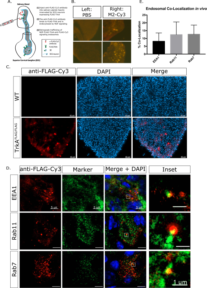
Figure 7.
SEs are diversified in vivo. (A) SEs can be labelled in vivo by injection into the salivary glands, shown diagrammatically. (B) Specificity of SE labelling. Injection of M2-Cy3 into the right eye and PBS into the left eye shows exclusive signal in the ipsilateral (right) SCG but not the contralateral (left) SCG. (C) Bright antibody labelling is detected when M2-Cy3 is injected into the salivary gland of TrkAFLAG/FLAG animals, but no label is observed when M2-Cy3 is injected into WT (non-FLAG) animals. (D) Close up imaging of single cells shows partial co-localization of in vivo SEs in the SCG with EEA1, Rab11, and Rab7. Scale bar 5 µm, inset scale bar 1 µm. (E) Quantification of SE co-localization with Rab proteins (n = 30–40 cells from 2 independent experiments).

