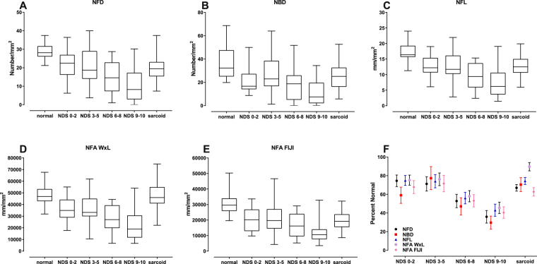
Figure 2.
Descriptive statistics of corneal confocal microscopy (CCM) variables (A–E) (median with interquartile range and maximum/minimum). In general, values of all variables decrease with increasing severity of neuropathy. The sarcoidosis group was generally similar to the NDS (0–2) and (3–5) groups for NFD, NBD, NFL, NFA FIJI, but diverged for NFA WxL. (F) Percent reduction from the corresponding means of normal subjects for each CCM variable (error bars: SEM) illustrates that small nerve fiber losses range from 80% to 40% with increasing severity of neuropathy. [NFD = nerve fiber density; NBD = main nerve branch density; NFL = nerve fiber length; NFA WxL = nerve fiber area determined by length times average width; NFA FIJI = nerve fiber area determined by the the total number of pixels within the nerve plexus].

