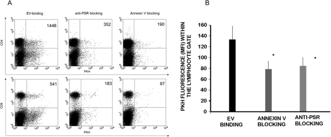
Figure 5.
Blocking the exofacial phosphatidylserine binding sites of embryo-derived EVs inhibits the binding of EVs to the lymphocytes. Prior to incubating embryo-derived EVs with anti-CD4 and anti-CD8 labelled splenocytes, we masked phosphatidylserine on the membranes of EVs by AnnexinV, or the phosphatidylserine receptors on the lymphocytes and checked the number of EVs binding CD4+ and CD8+ cells. Panel A: The upper right quadrants of representative dot plots show the PKH+/CD4+ or PKH+ /CD8+ double positive cells. These populations represent the number of lymphocytes which bound PKH-labelled EVs on their surface. Both anti-PSR antibody pretreatment of lymphocytes, and blocking of the exofacial phosphatidylserine on embryo-derived EVs by Annexin V partially inhibited the binding of EVs. Panel B: For comparing EV binding of lymphocytes with or without blocking, we used the PKH fluorescence intensity parameter (geometric mean channel values/MFI values) which reflects on the number of EVs per cell. PKH fluorescence was measured in the lymphocyte gate. Significantly lower (p = 0.05) PKH fluorescence was detected both after anti-PSR antibody pretreatment and Annexin V blocking. The bars represent the mean+/− SEM of 4 experiments.

