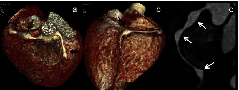
Figure 1. (a–c): CT Coronary angiography (CTCA) images (a & b - volume rendered images-VRT) and (c) curved maximum intensity projection (MIP) images of a 7 year male child in acute phase show two fusiform aneurysms in proximal right coronary artery (RCA) and one in distal RCA at its bifurcation into its posterior descending and postero-lateral branches (thin arrows).
Note fusiform aneurysm in mid left anterior descending artery (LAD) as well (a-thick arrow). 2D-transthoracic echocardiography could detect one aneurysm in proximal RCA and proximal LAD and missed two aneurysms in RCA.

