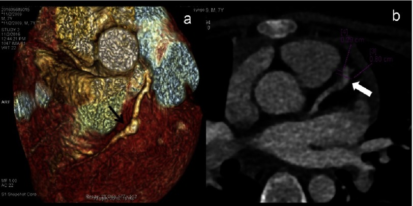
Figure 4. (a–b): CTCA VRT (a) axial (b) images of a 7 year male child in convalescent phase (4 years after diagnosis) show fusiform aneurysmal dilation (a - thin arrows) of proximal LAD; (b) shows a giant aneurysm with eccentric thrombus along the anterior wall (thick arrow).
2D-transthoracic echocardiography though could detect aneurysm but thrombus was missed.

