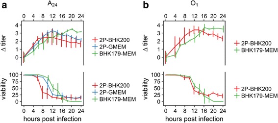
Fig. 3.

Virus infection kinetics on monolayer and suspension BHK cells. Virus infection kinetics were performed using the virus variants A24-2P to infect the suspension BHK-2P cell line, maintained in BHK200 (red line) or GMEM + 5% FCS (blue line) and A24–179 to infect the monolayer BHK179 cell line (green line) at an MOI = 0.1 (panel a). The virus variant O1-2P was used to infect the suspension BHK-2P cell line, maintained in BHK200 (red line), and the virus variant O1–179 was used to infect the monolayer BHK179 cell line (green line) under the same conditions (panel b) as described for serotype A. All preparations were sampled for the first time after 4 h and every 2 h thereafter. CPE and cell viability are given in percent (%). Titers are shown in log10 TCID50 relative to time 0
