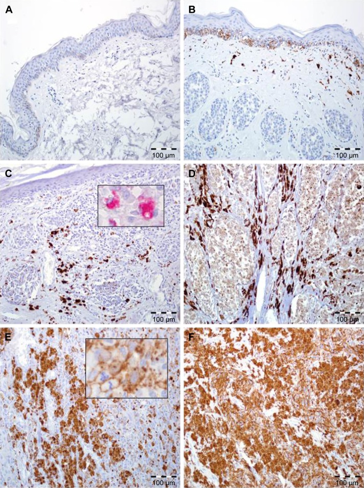
Figure 1.
FOXP1 expression in skin melanoma patients.
Notes: (A) Normal skin without FOXP1 immunoreactivity (200×, hematoxylin). (B) Intermediate expression of FOXP1 in the basal epidermal cells and normal melanocytes with lack of immunoreactivity in nests of benign melanocytes in intradermal skin nevus. Note the moderate reaction in macrophages and fibroblasts (200×, hematoxylin). (C) Diffuse, strong FOXP1 reactivity in stromal cells (predominantly in tumor-associated macrophages) with lack of expression in malignant melanocytes; inset: immunohistochemical reaction (red chromogen) with anti-CD68 antibody (200×, 600×, hematoxylin). (D) Enhanced FOXP1 expression in stromal cells with low immunoreactivity in melanoma cells (200×, hematoxylin). (E) High FOXP1 expression in neoplastic melanocytes, higher magnification: FOXP1-positive melanoma cells with atypical mitotic figure (200×, 600×, hematoxylin). (F) Diffuse, strong FOXP1 reactivity in melanoma cells with lack of expression in stromal compartment (200×, hematoxylin).

