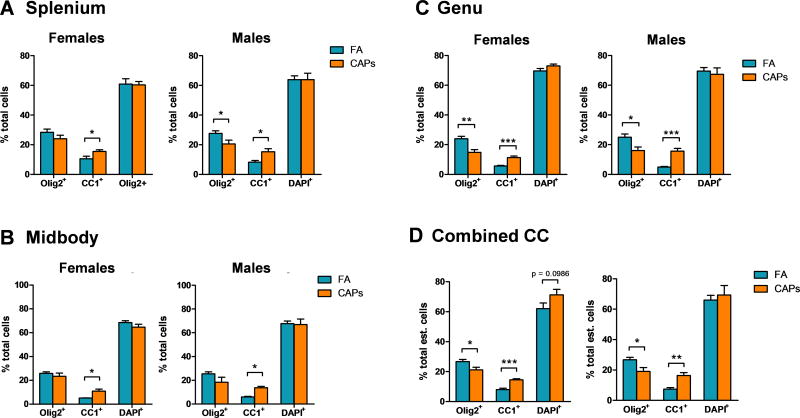
Figure 2. CAPs exposure proportionally shifts CC oligodendrocytes towards a more mature phenotype at PND11–15.
Stereological cell counts are expressed as a percent of total counted cells by CC sub-region and total CC. A) The proportion of mature CC-1+ oligodendrocytes is significantly increased in the splenium of males and females. In the male splenium, CAPs exposure significantly decreased the proportion of Olig2+ cells. B) In the midbody, only the proportion of CC-1+ cells was significantly increased by CAPs exposure in both sexes. C) Both the male and female genu experience a significant proportional decrease in Olig2 staining and a significant increase in CC-1 staining. D) Across the entire CC, there is a significant decrease in Olig2 staining with a corresponding increase in CC-1 staining. Additionally, there is a trending increase in the proportion of DAPI+ cells across the CC of CAPs-treated females. Data represent mean ± SEM of estimates across 3 serial tissue sections analyzed by one-way ANOVA (N = 6–11/group). *p < 0.05, **p < 0.01, ***p < 0.0001.

