Figure 2.

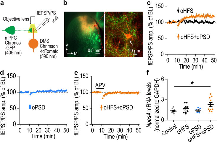
oPSD facilitated corticostriatal LTP induction in the DMS. (a) Schematic illustration of selective pre- and post-synaptic stimulation of DMS corticostriatal synapses using dual-channel optogenetics. AAV-Chronos-GFP was infused into the mPFC and AAV-Chrimson-tdTomato into the DMS of rats. Chronos and Chrimson were activated by 405- and 590-nm light, respectively. (b) Confocal fluorescent images showing Chronos-GFP-expressing mPFC fibers (green) and Chrimson-tdTomato-expressing neurons (red) in the DMS. Images shown in b is representative of 3 experiments of 3 rats. (c) Pairing oHFS with oPSD produced robust LTP in the DMS (119.06 ± 4.69% of baseline [BL], t(6) = −4.07, P = 0.0066; n = 7 slices, 4 rats). oHFS alone did not alter fEPSP/PS (99.60 ± 3.12% of BL, t(7) = 0.13, P = 0.90; n = 8 slices, 5 rats). (d) oPSD alone did not induce LTP (104.55 ± 3.15% of BL, t(6) = −1.44, P = 0.20; n = 7 slices, 3 rats). (e) Dual-channel optogenetic induction of LTP was blocked by APV (98.06 ± 3.27% of BL; t(8) = 0.59, P = 0.57; n = 9 slices, 5 rats). (f) Npas4 mRNA levels were significantly increased following paired oHFS+oPSD, but not after oHFS or oPSD only. F(3,30) = 3.86, P = 0.019; *P < 0.05; n = 10 (Control), 9 (oHFS), 5 (oPSD), and 10 (oHFS+oPSD) rats. Two-sided paired t test for c-e; one-way ANOVA followed by SNK test for f. Data are presented as mean ± s.e.m.
