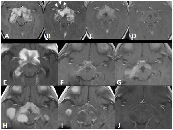
Fig. 1.
Axial contrast-enhanced T1-weighted MRIs in patients 1 (A–D) and 2 (E–J). Patient 1: (A) At baseline, a heterogeneously enhancing mass measuring 7.6×6.4×4.2 cm3 involved the optic chiasm, hypothalamus, and postchiasmatic optic pathways. (B) Six months after chemotherapy was initiated, overall enhancement in mass was slightly decreased and the tumor measured 7.3×6.2×4 cm3; however, a new anterior cystic/solid extension was apparent (arrowheads). (C) Eight weeks after initiating vemurafenib therapy, the lesion had decreased in size and enhancement. (D) At the 5-month follow-up, the tumor further decreased in size and enhancement and measured 5.9×4.4×1.8 cm3. Patient 2: (E) The baseline image shows enhancement and enlargement of the optic chiasm and hypothalamus, with tumor dimensions of 5.6×4.3×2.7 cm3. Also, the right optic pathway was larger than the left optic pathway. (F) After 13 months of chemotherapy, these features slowly improved. (G) At 15 months after initiating chemotherapy, the tumor had increased in size and enhancement. (H) Further progression was obvious after 1 year of weekly vinblastine therapy, and the tumor measured 5.1×9.4×4.5 cm3. (I) After 2 months of vemurafenib, there was dramatic improvement in the lesion. (J) At the 6-month follow-up, the tumor showed continued improvement and measured 4.1×9.2×4 cm3.

